
Surface-Enhanced Raman Scattering을 활용한 DNA·RNA 검출 기술의 원리
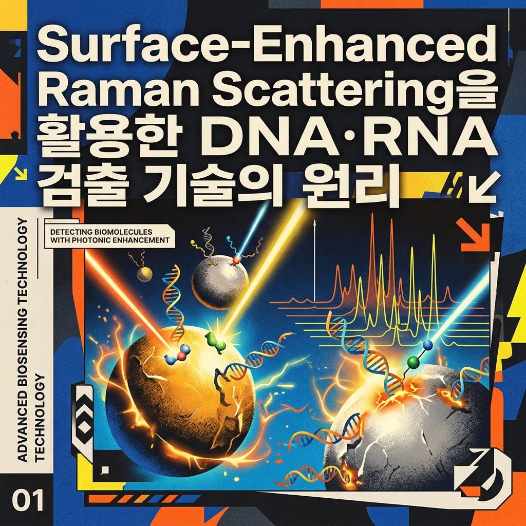
Surface-Enhanced Raman Scattering(SERS)은 금이나 은 같은 금속 나노구조 주변에서 Raman scattering 신호가 크게 증가하는 현상을 이용합니다. 핵산 probe와 Raman reporter를 이 영역에 배치하면 DNA·RNA hybridization을 고유한 spectral signal로 변환할 수 있습니다.
Raman Scattering부터 이해해야 하는 이유
빛을 물질에 조사하면 대부분은 입사광과 같은 에너지를 가진 상태로 산란됩니다.
이를 Rayleigh scattering이라고 합니다.
하지만 매우 적은 일부 photon은 molecule의 vibrational state와 energy를 주고받은 뒤 입사광과 다른 energy를 가지고 산란될 수 있습니다.
이 현상이 Raman scattering입니다.
입사광과 산란광 사이의 energy difference를 Raman shift로 측정하면 molecule의 chemical bond와 molecular vibration에 관한 정보를 얻을 수 있습니다.
따라서 Raman spectrum에는 여러 peak가 나타날 수 있고 각 peak의 위치는 특정 molecular vibration과 연결될 수 있습니다.
이러한 특성 때문에 Raman spectroscopy는 흔히 molecular fingerprint를 제공할 수 있는 분석법으로 설명됩니다.
일반 Raman Signal은 매우 약하다
Raman spectroscopy의 가장 큰 한계 가운데 하나는 scattering efficiency가 낮다는 점입니다.
Sample에 많은 photon을 조사하더라도 Raman scattering으로 검출되는 photon의 비율은 매우 작습니다.
특히 낮은 농도의 biomarker를 측정해야 하는 molecular diagnostics에서는 이 약한 signal이 문제가 됩니다.
DNA나 RNA가 극미량 존재하는 상황에서는 일반적인 Raman signal만으로 충분한 sensitivity를 확보하기 어려울 수 있습니다.
이 문제를 해결하기 위해 금속 nanostructure를 이용해 Raman signal을 크게 높이는 방법이 연구됐습니다.
그것이 Surface-Enhanced Raman Scattering입니다.
SERS에서는 왜 금과 은을 사용할까?
SERS substrate에는 gold(Au)와 silver(Ag) nanostructure가 널리 사용됩니다.
이러한 noble metal의 conduction electron은 특정 조건에서 incident electromagnetic field와 상호작용합니다.
Nanoparticle surface의 free electron이 집단적으로 진동하면서 localized surface plasmon resonance(LSPR)가 발생할 수 있습니다.
적절한 wavelength의 light가 조사되면 metal surface 주변에 강한 local electromagnetic field가 형성됩니다.
Raman-active molecule이 이 영역에 위치하면 molecule이 경험하는 electromagnetic field가 커집니다.
그 결과 Raman scattering intensity가 크게 증가할 수 있습니다.
SERS의 핵심은 Local Electromagnetic Field다
SERS enhancement를 설명할 때 가장 중요한 mechanism은 electromagnetic enhancement입니다.
금속 nanostructure 주변에서 incident light에 의해 local field가 증폭됩니다.
Molecule이 이 강한 field와 상호작용하면서 Raman excitation과 scattering이 모두 강화될 수 있습니다.
SERS에는 molecule과 metal surface 사이의 charge-transfer interaction 등에 의한 chemical enhancement도 기여할 수 있습니다.
하지만 일반적으로 큰 enhancement의 주요 원인으로 electromagnetic mechanism이 중요하게 다뤄집니다.
따라서 SERS sensor를 설계할 때는 reporter molecule의 종류뿐 아니라 금속 nanostructure의 geometry가 매우 중요합니다.
Hot Spot이란 무엇일까?
SERS에서 자주 등장하는 개념이 hot spot입니다.
두 개의 metal nanoparticle이 매우 가까이 위치하면 particle 사이의 좁은 gap에서 electromagnetic field가 매우 강해질 수 있습니다.
이처럼 특별히 강한 field가 형성되는 nanoscale region을 hot spot이라고 합니다.
Raman reporter가 hot spot 안에 위치하면 particle에서 멀리 떨어져 있을 때보다 훨씬 강한 SERS signal을 만들 수 있습니다.
따라서
Nanoparticle Aggregation
→ Nanoscale Gap Formation
→ Strong Local Field
→ Enhanced Raman Signal
을 이용한 biosensor를 설계할 수 있습니다.
그러나 nanoparticle을 무작정 많이 응집시키는 것이 좋은 것은 아닙니다.
불규칙한 aggregation은 hot spot의 위치와 수를 일정하게 만들기 어렵게 해 measurement reproducibility를 떨어뜨릴 수 있습니다.
SERS로 DNA 자체를 직접 측정할 수 있을까?
가능한 접근이지만 해석에 주의해야 합니다.
DNA와 RNA의 base, sugar와 phosphate group은 각각 Raman-active molecular vibration을 가집니다.
따라서 nucleic acid가 metal surface 가까이에 존재하면 직접적인 Raman spectrum을 측정하는 연구가 가능합니다.
이를 label-free SERS detection으로 접근할 수 있습니다.
그러나 실제 biosensor에서는 target concentration이 낮고 biological sample에 많은 다른 molecule이 존재합니다.
여러 spectral peak가 겹칠 수도 있습니다.
따라서 target nucleic acid 자체의 Raman spectrum을 직접 분석하는 방법 외에도 Raman reporter를 이용한 간접 검출 방식이 많이 사용됩니다.
Raman Reporter란 무엇일까?
Raman reporter는 강하고 특징적인 Raman peak를 생성하도록 선택한 molecule입니다.
이 reporter를 gold 또는 silver nanoparticle에 부착할 수 있습니다.
그리고 nanoparticle 표면에 target DNA와 결합하는 probe를 함께 배치할 수 있습니다.
Target nucleic acid가 존재하면 reporter-containing nanoparticle의 위치나 aggregation state가 변하도록 설계합니다.
그 결과 SERS intensity가 증가하거나 감소할 수 있습니다.
즉, 실제로 측정하는 spectrum은 target DNA 자체보다 target recognition에 의해 상태가 변한 Raman reporter에서 나올 수 있습니다.
SERS Tag는 어떻게 만들까?
SERS biosensor에서 사용하는 nanoparticle probe는 흔히 여러 기능을 하나의 particle에 결합합니다.
개념적으로
Metal Nanoparticle
- Raman Reporter
- Target Recognition Probe
로 구성할 수 있습니다.
Metal nanoparticle은 electromagnetic enhancement를 제공합니다.
Raman reporter는 분석에 사용할 spectral fingerprint를 만듭니다.
DNA probe는 target specificity를 제공합니다.
이 세 요소를 결합하면 target sequence를 Raman signal로 변환하는 SERS tag를 만들 수 있습니다.
Sandwich Assay와 결합하는 방법
DNA target을 검출하는 대표적인 방식 가운데 하나가 sandwich hybridization입니다.
첫 번째 capture probe는 substrate 또는 nanoparticle에 고정합니다.
Target DNA가 capture probe와 hybridization합니다.
그 다음 target의 다른 영역을 인식하는 detection probe가 결합합니다.
Detection probe에 SERS nanoparticle tag가 연결되어 있다면 target이 존재할 때 Raman reporter가 detection region에 축적됩니다.
즉,
Capture Probe
→ Target DNA
→ SERS Detection Probe
구조가 만들어집니다.
Target 농도가 높을수록 capture되는 SERS tag 수가 증가하도록 설계하면 Raman peak intensity를 이용해 target을 정량할 수 있습니다.
두 Nanoparticle 사이를 Target이 연결하도록 만들 수도 있다
SERS의 hot spot 특성을 직접 이용하는 방식도 있습니다.
서로 다른 두 nanoparticle에 target의 서로 다른 영역을 인식하는 DNA probe를 부착합니다.
Target DNA가 없다면 particle이 떨어져 있습니다.
Target이 존재하면 두 probe에 동시에 hybridization하면서 nanoparticle을 가까이 연결할 수 있습니다.
Particle 사이에 nanoscale gap이 형성되면 strong hot spot이 만들어질 수 있습니다.
따라서
Target 없음
→ Particles Separated
→ Weak SERS
반면
Target 존재
→ DNA-Mediated Particle Assembly
→ Hot Spot Formation
→ Strong SERS
와 같은 signal-on assay를 만들 수 있습니다.
Aggregation-Based Sensor의 어려움
Target-dependent nanoparticle aggregation은 직관적이지만 정량 분석에서는 문제가 발생할 수 있습니다.
Particle aggregation은 항상 동일한 크기와 구조로 형성되지 않습니다.
어떤 aggregate에는 강한 hot spot이 많이 생길 수 있고 다른 aggregate에는 적게 생길 수 있습니다.
Laser spot이 어느 위치를 측정했는지에 따라서도 intensity가 달라질 수 있습니다.
따라서 동일한 target concentration에서도 signal variation이 커질 수 있습니다.
이 문제 때문에 정교하게 제작한 nanostructure나 내부 표준을 사용하는 SERS assay가 연구됩니다.
Gold Nanostar가 사용되는 이유
완전히 둥근 nanoparticle만 SERS substrate로 사용되는 것은 아닙니다.
Gold nanostar처럼 sharp tip을 가진 nanostructure도 이용됩니다.
Sharp metallic feature 주변에서는 electromagnetic field가 집중될 수 있습니다.
이를 흔히 lightning-rod effect와 연결해 설명합니다.
Nanostar의 tip과 다른 nanoparticle 사이의 gap을 조절하면 강한 hot spot을 만들 수도 있습니다.
Nanorod, nanocube, nanosphere, core-shell particle 등 다양한 geometry가 연구되는 이유도 local electromagnetic field를 효과적으로 제어하기 위해서입니다.
Gold와 Silver는 어떤 차이가 있을까?
Silver nanostructure는 매우 강한 SERS enhancement를 제공할 수 있어 널리 연구됩니다.
하지만 surface oxidation과 chemical stability 문제가 고려될 수 있습니다.
Gold는 비교적 높은 chemical stability와 biomolecule functionalization의 편의성 때문에 biosensor에서 많이 사용됩니다.
특히 thiol-modified DNA를 gold surface에 고정하기 쉽습니다.
따라서 어느 metal이 항상 우수하다고 말하기보다 excitation wavelength, required enhancement, surface chemistry와 stability를 고려해 선택해야 합니다.
miRNA도 SERS로 검출할 수 있다
miRNA는 약 18~24 nucleotide로 짧기 때문에 SERS biosensor에서도 특별한 probe design이 필요합니다.
Target miRNA가 nanoparticle aggregation을 유도하도록 만들 수 있습니다.
또는 miRNA가 hairpin probe를 열면서 Raman reporter를 hot spot에 이동시키도록 설계할 수도 있습니다.
Target recycling reaction을 추가해 하나의 miRNA가 여러 SERS tag의 상태를 변화시키도록 만들 수도 있습니다.
즉,
miRNA Recognition
→ Molecular Structure Change
→ Nanoparticle Distance Change
→ SERS Intensity Change
로 target을 측정할 수 있습니다.
Molecular Beacon과 SERS를 연결하면
Molecular beacon에서는 target이 hairpin을 열면서 fluorophore와 quencher 사이의 거리가 변합니다.
비슷한 structure-switching concept을 SERS에 적용할 수도 있습니다.
DNA hairpin 한쪽에 metal surface와 결합하는 영역을 두고 Raman reporter의 위치가 target binding에 따라 변하도록 설계할 수 있습니다.
Hairpin이 닫혀 있을 때 reporter가 metal surface 또는 hot spot 가까이에 있다면 강한 SERS signal이 나타날 수 있습니다.
Target hybridization으로 hairpin이 열리면서 reporter가 멀어지면 signal이 감소할 수 있습니다.
반대 방향의 signal design도 가능합니다.
따라서 SERS는 단순히 nanoparticle aggregation만 측정하는 기술이 아닙니다.
DNAzyme과 SERS를 결합할 수도 있다
Target recognition 후 DNAzyme이 활성화되도록 만들고 substrate cleavage를 nanoparticle assembly 변화와 연결할 수 있습니다.
예를 들어 DNAzyme cleavage 전에는 두 nanoparticle이 연결되어 strong hot spot을 유지하고 있다고 가정할 수 있습니다.
Target-dependent catalytic reaction으로 linker가 절단되면 particle이 분리됩니다.
SERS intensity가 감소합니다.
반대로 cleavage가 nanoparticle assembly를 유도하도록 설계하면 signal-on 방식도 가능합니다.
따라서 SERS는 다양한 nucleic acid catalytic circuit의 최종 readout으로 사용할 수 있습니다.
HCR과 결합하면 많은 SERS Tag를 모을 수 있다
Hybridization Chain Reaction은 긴 DNA assembly를 형성합니다.
Target recognition으로 HCR이 시작되면 반복적인 H1-H2 structure가 생성됩니다.
HCR product에 SERS tag가 결합할 수 있는 반복 sequence를 넣을 수 있습니다.
그러면 하나의 target recognition event에서 여러 SERS nanoparticle이 모집될 수 있습니다.
즉,
Target
→ HCR
→ Long DNA Assembly
→ Multiple SERS Tag Binding
→ Enhanced Raman Signal
로 amplification을 구현할 수 있습니다.
이 경우 signal amplification의 첫 번째 원인은 HCR이며 SERS enhancement는 optical readout sensitivity를 추가하는 역할을 합니다.
RCA와 SERS의 결합
Rolling Circle Amplification 역시 SERS와 연결하기 좋은 nucleic acid amplification method입니다.
RCA product에는 동일한 sequence가 반복됩니다.
각 repeat에 complementary한 SERS tag를 결합시키면 하나의 RCA product에 많은 nanoparticle reporter를 모집할 수 있습니다.
또는 RCA product 자체를 이용해 nanoparticle aggregation을 유도할 수도 있습니다.
따라서
Sequence Recognition
→ Padlock Probe Ligation
→ RCA
→ Repeated Binding Sites
→ SERS Tag Accumulation
과 같은 assay architecture를 만들 수 있습니다.
CRISPR와 SERS도 결합할 수 있을까?
CRISPR nuclease의 target-dependent cleavage를 nanoparticle assembly와 연결할 수 있습니다.
예를 들어 Cas12의 collateral cleavage substrate를 두 SERS nanoparticle 사이의 DNA linker로 사용할 수 있습니다.
Target이 없으면 linker가 유지되어 particle distance가 일정하게 유지됩니다.
Target DNA가 존재하면 Cas12가 활성화되고 linker가 절단될 수 있습니다.
그 결과 nanoparticle distance가 변하면서 SERS signal이 달라집니다.
이 경우
CRISPR → Sequence-Specific Recognition
DNA Linker → Signal Transducer
SERS → Optical Readout
역할을 담당합니다.
SERS 자체는 핵산 증폭법이 아니다
매우 강한 Raman enhancement가 나타난다고 해서 target DNA가 증폭되는 것은 아닙니다.
SERS는 optical signal enhancement 기술입니다.
PCR처럼 target copy를 늘리지 않습니다.
RCA처럼 새로운 DNA product를 합성하지도 않습니다.
CHA처럼 target recycling을 자동으로 수행하는 것도 아닙니다.
따라서 SERS assay에서 amplification을 논할 때는
Target Amplification
Molecular Signal Amplification
Raman Enhancement
를 구분해야 합니다.
SERS는 주로 세 번째 역할을 담당합니다.
Raman Peak의 위치와 세기는 다른 정보다
SERS spectrum을 해석할 때 peak position과 intensity를 구분해야 합니다.
Peak position은 reporter의 molecular vibration과 관련된 spectral identity를 제공합니다.
Intensity는 reporter의 양, hot spot 위치, particle geometry와 laser condition 등에 영향을 받습니다.
따라서 특정 Raman shift에서 peak가 나타난다는 것은 reporter identity를 확인하는 데 활용할 수 있고 peak intensity 변화는 target quantification에 사용할 수 있습니다.
하지만 intensity는 여러 physical factor의 영향을 받기 때문에 calibration이 중요합니다.
Multiplex Detection에서 SERS가 주목받는 이유
Fluorescence multiplexing에서는 fluorophore의 broad emission spectrum이 서로 겹치는 문제가 발생할 수 있습니다.
Raman spectrum의 peak는 상대적으로 좁게 나타날 수 있습니다.
서로 다른 Raman reporter가 구별되는 spectral fingerprint를 갖도록 선택하면 하나의 laser excitation 아래에서 여러 reporter를 구분하는 전략이 가능합니다.
예를 들어 Target A에는 Reporter A를 연결하고 Target B에는 Reporter B를 연결할 수 있습니다.
Spectrum에서 서로 다른 Raman peak를 분석하면 두 target을 동시에 구별할 수 있습니다.
이 때문에 SERS는 multiplex molecular diagnostics 연구에서 관심을 받습니다.
Raman Reporter를 많이 넣으면 항상 좋은 것일까?
Reporter 수가 증가하면 signal이 증가할 가능성이 있습니다.
하지만 nanoparticle surface에 reporter를 지나치게 많이 배치하면 recognition probe의 접근성을 방해할 수 있습니다.
Surface chemistry가 변하면서 particle stability에 영향을 줄 수도 있습니다.
또한 SERS signal은 reporter molecule의 단순한 총량만으로 결정되지 않습니다.
Reporter가 실제로 strong electromagnetic hot spot에 얼마나 가까이 위치하는지가 매우 중요합니다.
따라서 Reporter Number와 Reporter Position을 함께 고려해야 합니다.
Hot Spot의 재현성이 가장 어려운 문제 중 하나다
SERS 연구에서 매우 높은 enhancement factor가 보고될 수 있지만 모든 measurement point가 동일한 enhancement를 제공하는 것은 아닙니다.
Nanoparticle 사이의 gap이 조금만 달라져도 local electromagnetic field가 크게 변할 수 있습니다.
Particle size와 shape variation도 영향을 줍니다.
따라서 random nanoparticle aggregation에 의존하는 assay에서는 spot-to-spot variation이 커질 수 있습니다.
실제 quantitative diagnostic platform으로 발전하려면 reproducible SERS substrate fabrication이 중요합니다.
Internal Standard를 사용하는 이유
Measurement variation을 줄이기 위해 internal standard를 사용할 수 있습니다.
Target-dependent reporter signal만 단독으로 측정하는 대신 target과 관계없이 일정하게 유지되는 reference Raman peak를 함께 측정합니다.
그 다음 두 peak의 intensity ratio를 계산합니다.
예를 들어
I_target / I_reference
형태로 정규화할 수 있습니다.
이러한 ratiometric approach는 laser intensity 변화, substrate heterogeneity와 measurement condition의 일부 영향을 보정하는 데 도움이 될 수 있습니다.
Single-Nucleotide Mutation을 구별할 수 있을까?
SERS 자체가 sequence specificity를 제공하는 것은 아닙니다.
Target discrimination은 DNA probe나 upstream molecular reaction이 담당합니다.
완전 상보적인 target만 안정적인 nanoparticle assembly를 만들도록 probe를 설계할 수 있습니다.
Padlock probe ligation을 이용해 single-base difference를 구별한 뒤 RCA-SERS로 signal을 읽을 수도 있습니다.
CRISPR recognition을 upstream에 사용할 수도 있습니다.
따라서 SNV detection에서는
Sequence Recognition Module
과
SERS Readout Module
을 구분해서 평가해야 합니다.
Label-Free SERS의 장점과 어려움
Label-free detection에서는 별도의 Raman reporter를 붙이지 않고 target molecule 자체의 spectrum을 분석합니다.
Reporter synthesis와 labeling 과정이 줄어든다는 장점이 있습니다.
또한 molecule 자체의 vibrational information을 얻을 수 있습니다.
하지만 biological sample에는 수많은 Raman-active molecule이 함께 존재합니다.
DNA base 사이에서도 spectral feature가 일부 겹칠 수 있습니다.
Target concentration이 낮다면 원하는 spectrum을 background에서 분리하기 더욱 어렵습니다.
따라서 label-free SERS에서는 substrate reproducibility뿐 아니라 spectral data analysis도 중요합니다.
Machine Learning과 결합하는 이유
복잡한 SERS spectrum에는 여러 peak와 intensity pattern이 존재할 수 있습니다.
단일 peak 하나만으로 sample을 분류하기 어려운 경우 전체 spectral pattern을 이용할 수 있습니다.
Principal component analysis 같은 multivariate analysis나 machine-learning algorithm을 적용해 여러 sample group을 분류하는 연구가 가능합니다.
하지만 algorithm의 높은 classification accuracy가 자동으로 임상적 유효성을 의미하지는 않습니다.
Training dataset 규모, independent validation, batch effect와 overfitting을 함께 확인해야 합니다.
실제 혈액에서는 어떤 문제가 생길까?
Blood, serum과 plasma에는 protein, lipid와 다양한 metabolite가 존재합니다.
이들이 metal nanoparticle surface에 흡착하면 protein corona가 형성될 수 있습니다.
그 결과 DNA probe가 target에 접근하기 어려워질 수 있습니다.
Nanoparticle aggregation behavior도 변할 수 있습니다.
또한 biological molecule 자체에서 Raman background가 발생할 수 있습니다.
따라서 실제 liquid biopsy sample에 SERS sensor를 적용하려면 target extraction, surface blocking과 antifouling strategy가 필요할 수 있습니다.
Nanoparticle Stability도 중요하다
Colloidal SERS assay에서는 nanoparticle이 일정한 상태로 유지되어야 합니다.
Salt concentration이 높아지면 nanoparticle이 nonspecific aggregation을 일으킬 수 있습니다.
Biological sample의 protein도 particle surface chemistry에 영향을 줄 수 있습니다.
Target 때문에 발생한 aggregation과 sample matrix 때문에 발생한 aggregation을 구분하지 못하면 false signal이 나타날 수 있습니다.
따라서 nanoparticle-based assay에서는 colloidal stability 자체가 analytical specificity의 일부가 됩니다.
SERS 핵산 검출의 기본 구조를 정리하면
Target-induced hot spot formation sensor를 예로 들 수 있습니다.
Target이 없는 경우
Probe-Functionalized Nanoparticle A
Probe-Functionalized Nanoparticle B
→ Particle Separation
→ Weak Electromagnetic Coupling
→ Low SERS Signal
Target이 존재하는 경우
Target DNA Hybridization
→ Nanoparticle A-Target-B Assembly
→ Nanoscale Gap Formation
→ Electromagnetic Hot Spot
→ Raman Reporter Enhancement
→ High SERS Signal
실제 assay에서는 반대로 target이 particle을 분리해 signal을 낮추도록 설계할 수도 있습니다.
앞선 ECL과 비교하면 무엇이 다를까?
| 구분 | ECL | SERS |
|---|---|---|
| 기본 현상 | 전기화학적으로 유도된 발광 | 표면 증강 Raman scattering |
| 주요 구조 | Electrode + Luminophore | Plasmonic Nanostructure + Raman Reporter |
| 외부 광원 | 발광 excitation용 광원 불필요 | Raman excitation laser 필요 |
| 주요 측정값 | 발광 intensity | Raman spectrum |
| 분자 식별 | Reporter에 따라 가능 | 고유 Raman shift 활용 가능 |
| Multiplexing | Reporter 설계에 따라 가능 | 좁은 Raman peak 활용 가능 |
| 핵산 자체 증폭 | 하지 않음 | 하지 않음 |
두 기술 모두 nucleic acid recognition 결과를 물리적 신호로 읽는 readout platform이지만 signal이 만들어지는 mechanism은 완전히 다릅니다.
분자진단에서 SERS가 갖는 의미
분자진단에서는 target을 정확하게 인식하는 것만큼 매우 작은 molecular event를 어떻게 측정할 것인지가 중요합니다.
SERS는 plasmonic nanostructure 주변의 강한 local electromagnetic field를 이용해 Raman reporter의 약한 scattering을 크게 증가시키는 방법을 제공합니다.
여기에 DNA hybridization, aptamer, enzyme-assisted recycling, RCA, HCR 또는 CRISPR 같은 molecular recognition·amplification system을 연결할 수 있습니다.
또한 서로 다른 reporter의 좁은 Raman fingerprint를 이용하면 multiplex detection architecture를 구성할 가능성도 있습니다.
따라서 SERS의 가치는 단순히 ‘빛을 강하게 만드는 기술’보다 nanoscale plasmonic environment를 이용해 분자 인식 사건을 구별 가능한 spectral information으로 변환할 수 있다는 점에서 찾을 수 있습니다.
마치며
Surface-Enhanced Raman Scattering은 gold와 silver 같은 plasmonic metal nanostructure 주변에서 형성되는 강한 local electromagnetic field를 이용해 molecule의 Raman scattering을 증강하는 기술입니다.
핵산 바이오센서에서는
Target DNA·RNA Recognition
→ Nanoparticle Assembly 또는 Reporter Position 변화
→ Hot Spot 형성 또는 변화
→ Raman Scattering Enhancement
→ Characteristic Raman Spectrum Detection
으로 target 정보를 읽을 수 있습니다.
SERS 자체가 target DNA나 RNA를 복제하는 nucleic acid amplification technology는 아닙니다.
필요한 경우 PCR, RCA, HCR, target recycling 또는 CRISPR와 결합해 target recognition과 molecular amplification을 수행하고 SERS를 최종 readout으로 사용할 수 있습니다.
특히 SERS를 이해할 때 중요한 것은 nanoparticle이 단순히 Raman reporter를 운반한다는 점만이 아닙니다. 금속 나노구조 사이의 수 나노미터 수준의 geometry가 electromagnetic hot spot을 만들고, reporter가 그 공간의 어디에 위치하는지가 최종 Raman signal을 크게 좌우한다는 점이 핵심입니다.
따라서 SERS 기반 DNA·RNA 검출 기술은 nucleic acid sequence design뿐 아니라 plasmonic nanostructure, surface chemistry와 nanoscale distance control을 함께 다루는 분자진단 기술이라고 볼 수 있습니다.
※ 본 글은 SERS 기반 DNA·RNA 검출 원리와 바이오센서 연구를 설명하기 위한 교육 목적의 콘텐츠입니다. 연구 단계에서 보고된 검출한계나 분류 성능을 확립된 임상 진단검사의 성능으로 해석해서는 안 됩니다.



댓글 0
첫 댓글을 남겨보세요.